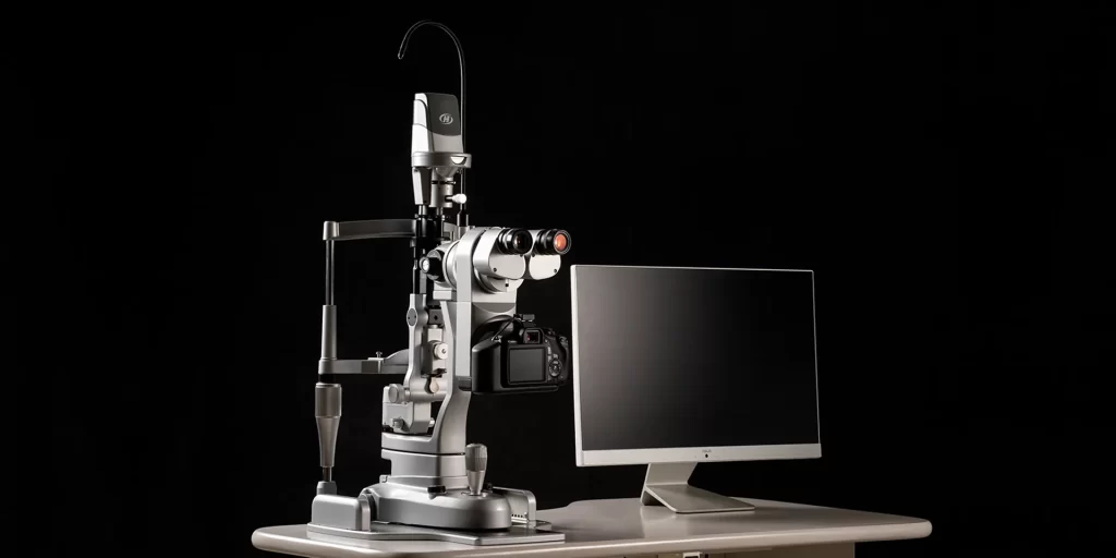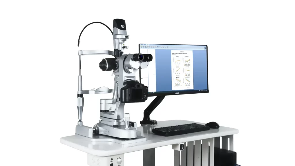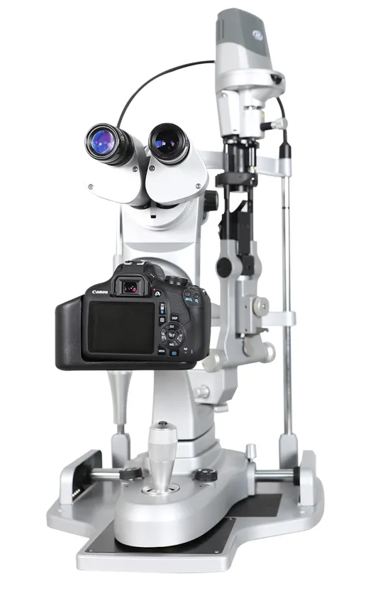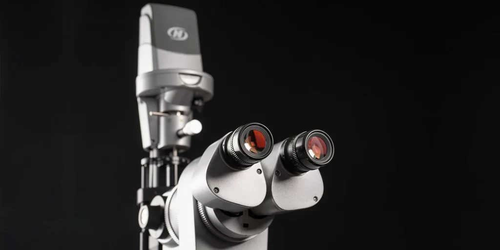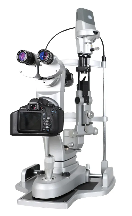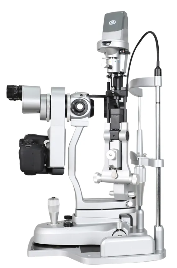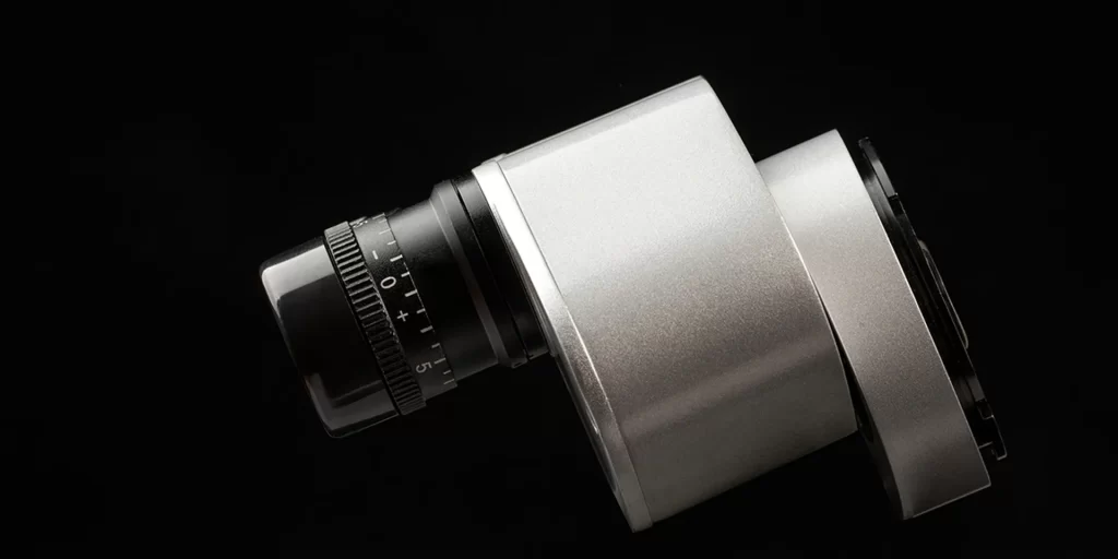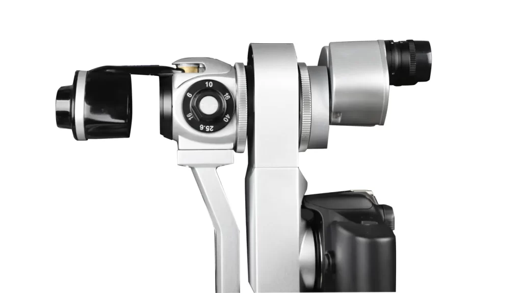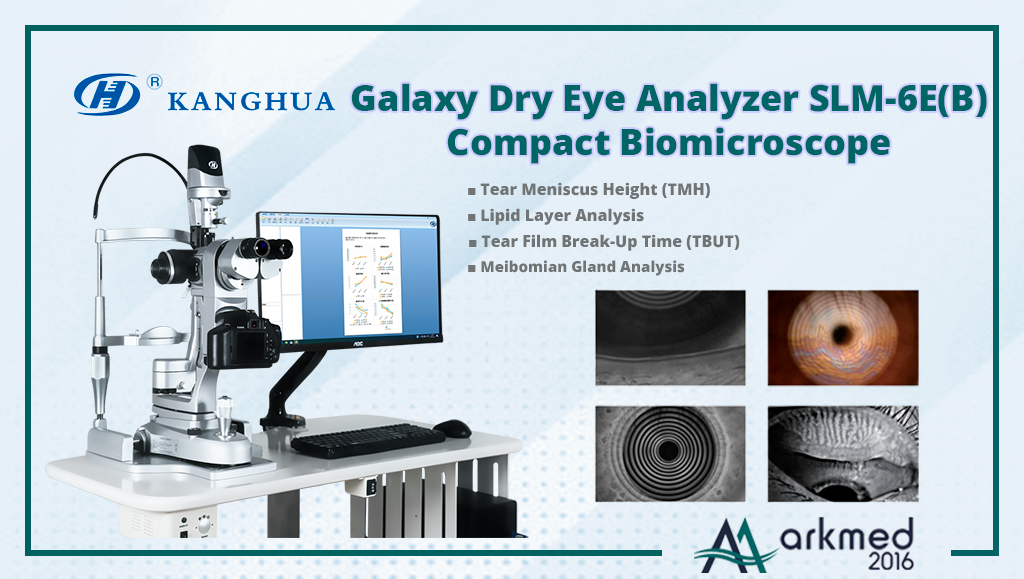
Kanghua Galaxy Dry Eye Analyzer SLM-6E(B) Compact Biomicroscope
Our classic ocular surface examination platform, built with advanced electronic control technology that combines excellent optical microscopy, intelligent light conversion technology, and high-resolution digital imaging, has been developed with a focus on our customers’ needs. When used alongside intelligent analysis software, it not only delivers high-resolution images and accurate analysis results for clinical dry eye examinations but also provides extremely clear cellular-level images and measurement data for more detailed study of ocular surface diseases, supporting academic research as well.
Comprehensive Analysis Report
Automatically generate reports based on examination results without manual input. Illustrated reports are easy to understand, making the process simpler. Clear and intuitive reports facilitate communication with patients.
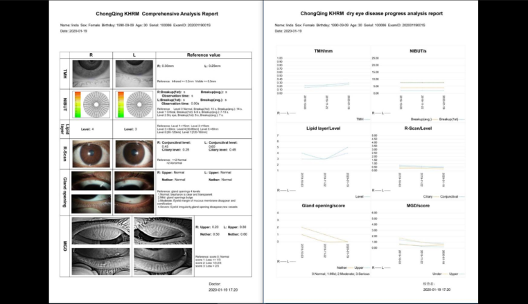
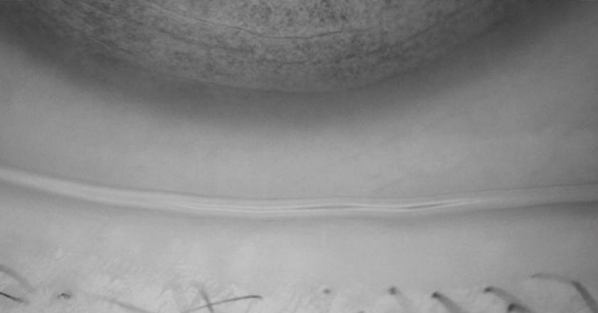
Tear Meniscus Height Measurement
Infrared light illumination is non-invasive and non-stimulating, and the tear secretion remains unchanged during measurement, ensuring high data accuracy for TMH.
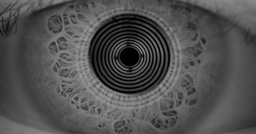
Auto NIBUT (Non-Invasive Break-Up Time)
Infrared light illumination differs from the stimulation caused by visible light to the subject’s eyes. This not only ensures the successful completion of the test but also preserves the natural dynamics of the tear film environment, making the test results accurate and reliable.
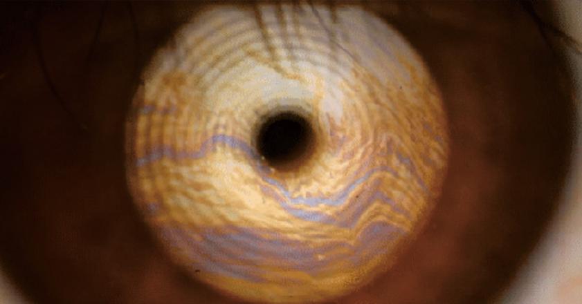
Lipid Layer Evaluation
The unique structural design of the dry eye device maximizes the use of light effects and, based on non-stimulating low-brightness illumination, presents the true color of the lipid layer, thereby ensuring the accuracy of lipid layer thickness assessment.
Meibomian Gland Expression
Meibomian gland imaging at the acinar level, combined with infrared imaging and optical magnification, provides detailed visualization of the acini, supporting clinicians in the immediate treatment of affected meibomian gland areas. Comparison with standard templates is performed to visually assess the degree of meibomian gland deficiency.
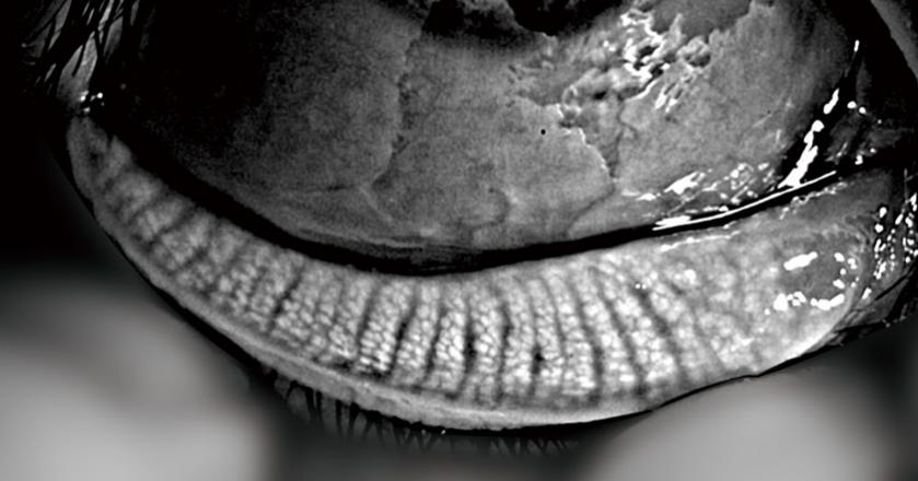
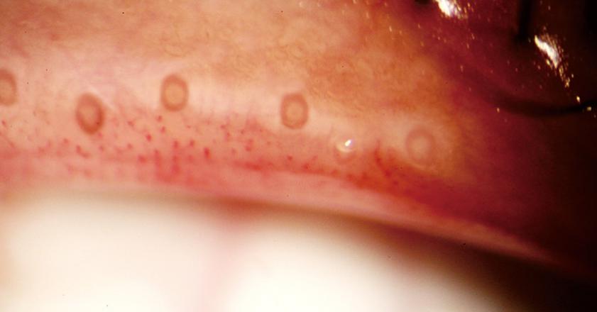
Eyelid Margin Analysis
High-resolution images of the eyelid margin can directly reflect the status of the meibomian gland openings, allowing easy identification of any blockages. Based on high-resolution imaging and comparison with standard templates, the degree of meibomian gland patency and changes in the eyelid margin can be visually assessed.
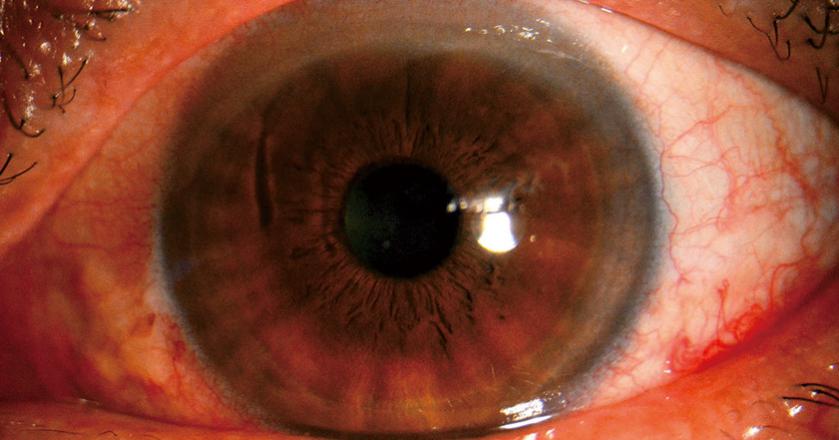
Redness Screening
Automatic quantification of the degree of ciliary conjunctival hyperemia to assess the severity of ocular surface inflammation.
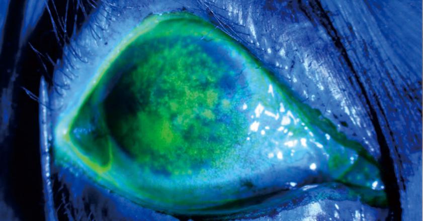
Corneal Staining
It reflects corneal epithelial integrity and assesses the severity of dry eye.